Neuro research

Prof Ben Loos
Head of department and research group leader
Quick links
Overview
The Neuro Research Group (NRG), headed by Prof Ben Loos, combines cell biology, cell physiology, microscopy and biochemistry approaches to dissect and investigate the relationship and role of protein degradation through macroautophagy and cell death susceptibility in neurodegeneration and gliomas.
The fine balance of proteostasis control, protein aggregation and proteotoxicity is the main focus point, due to its role in disease onset and progression. Autophagy is an essential process that allows cellular survival in stress conditions through the enhanced degradation of long-lived proteins. Since the vast majority of proteins in the cell are long-lived, its metabolic contribution is significant.
Dysfunction in autophagy is associated with numerous pathologies that are characterized by a shift in cell death susceptibility, such as neurodegenerative diseases or cancer. In order to achieve this, the lab focuses on macroautophagy (MA), chaperone mediated autophagy (CMA), cellular metabolism, mitochondrial morphology and function, tubulin and transport systems, the cytoskeleton and ATP consumption.
Central to our approach is a dynamic perspective on the cell’s function and its stress response, in context of its current intracellular and extracellular metabolic parameters. Our research findings in the past have demonstrated a clear relationship between the cell’s autophagic proficiency and apoptosis/necrosis onset, which demands for a dynamic and integrative approach.
Research focus areas
- Role of autophagy in disease pathogenesis
- Regulation and control of autophagic flux
- Microscopy
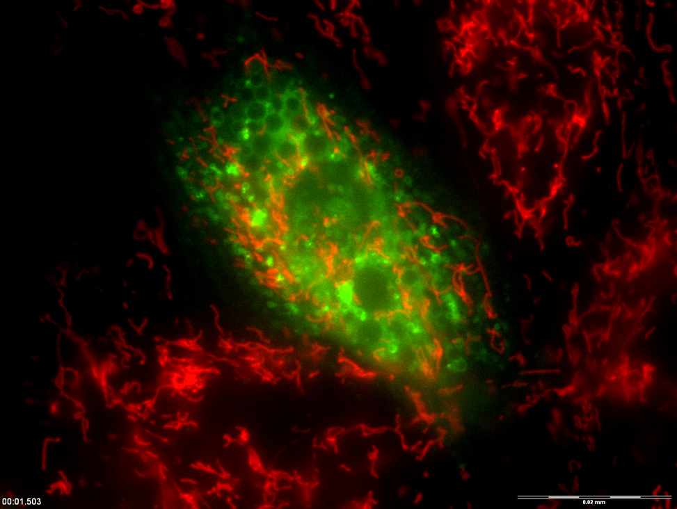
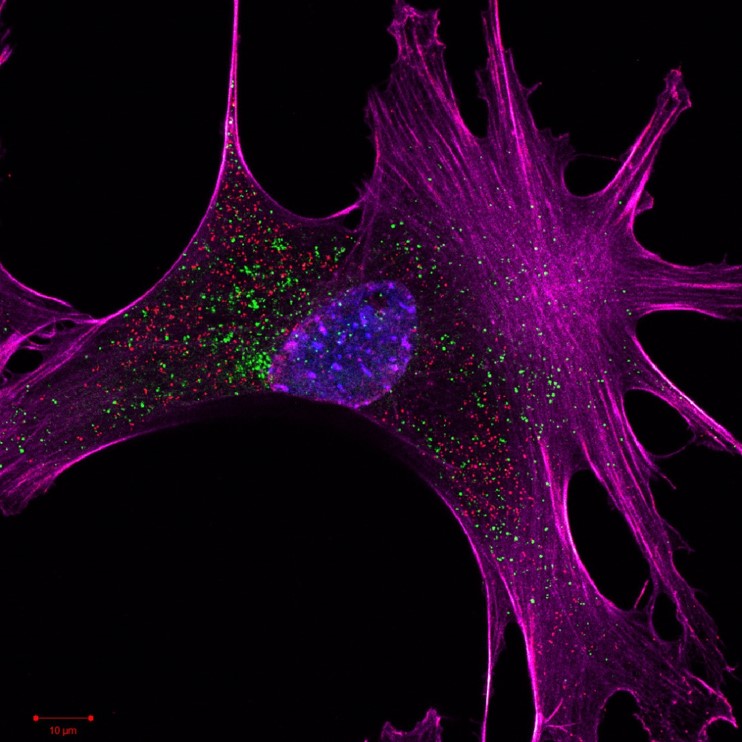
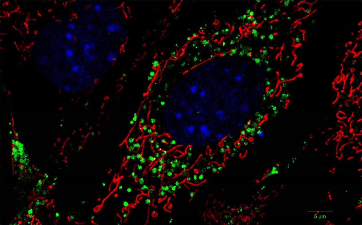
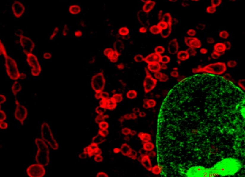
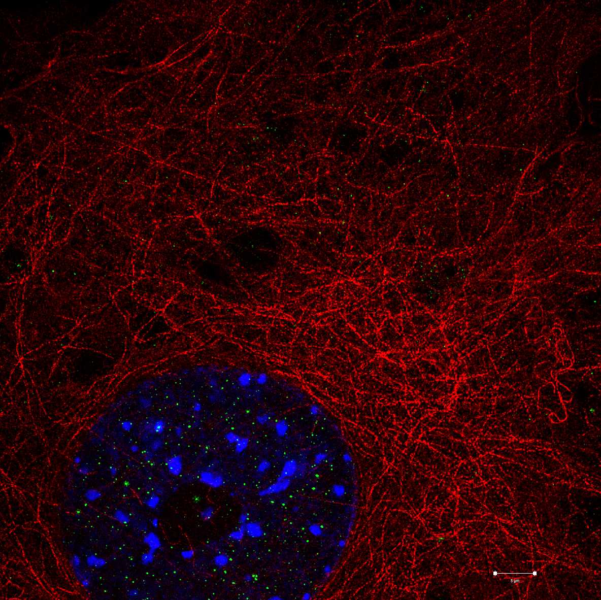
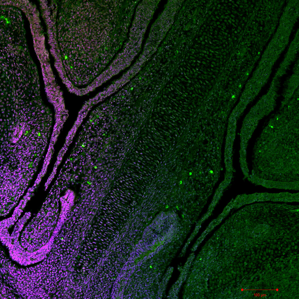
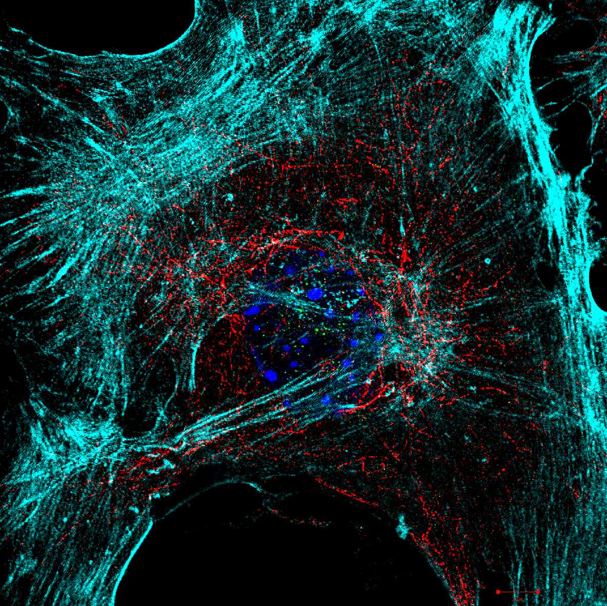
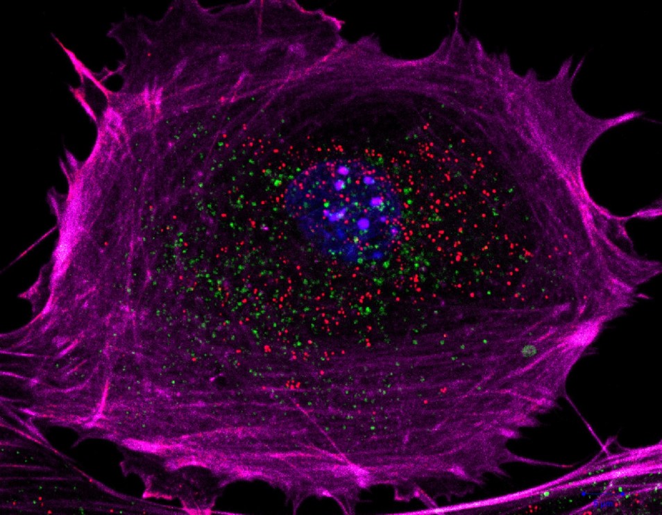
Microscopy
Confocal, Super resolution, SEM and more
In order to fully understand molecular behaviour within cells and tissues, we utilise microscopy techniques such as live cell imaging, fluorescence resonance energy transfer (FRET), fluorescence recovery after photobleaching (FRAP) or super-resolution structured illumination microscopy (SR-SIM) to generate data that can be utilized for statistical analysis.
SIM, which allows us to achieve a resolution of 80 nm, is thereby uniquely positioned to generate data that cannot be resolved through normal confocal imaging. Furthermore, we have recently started to utilize cutting edge techniques such Photoactivation Localization Microscopy (PALM) and Stochastic Optical Reconstruction Microscopy (STORM), to resolve structural detail up to 20 nm and Correlative Light and Electron Microscopy (CLEM) for immensely accurate localisation data.
By exploiting the functional aspects of fluorochromes, their spectral sensitivity in a defined environment (pH, voltage across membranes etc) we are able to tighten the gap between light microscopy and electron microscopy.