Confocal Microscopy
18 August 2022
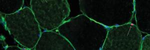
Confocal Microscopy
Sample: Muscle cross section
Instrument: ZEISS LSM780 CONFOCAL
The confocal microscope can be used for multi-colour fluorescence imaging. This is an image of a cross-section through a muscle biopsy, showing fast and slow twitch fibres (green), nuclei (blue) on the perimeter of each fibre and a few sparse satellite cells (pink).